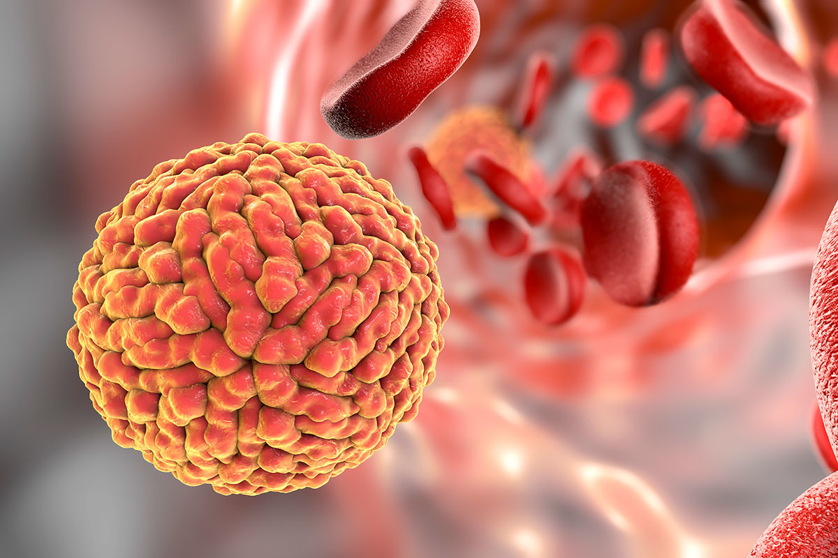
Novel way to track dengue infection
June 08, 2017 | Thursday | News
The hope is that non-invasive PET-FDG imaging can be used to transform the assessment of new dengue treatments in clinical trials so that infections may be more effectively treated in the clinic.
A team from Duke-NUS Medical School (Duke-NUS) and Singapore General Hospital (SGH) has found a new use for an 'old' technology in the field of infectious diseases research.
Using positron emission tomography (PET) paired with the glucose metabolism probe, fluorodeoxyglucose (FDG) as an imaging tool for dengue infection in mouse models, the team has potentially uncovered a novel and non-invasive way to track the infection in real-time and more accurately assess the effectiveness of new treatments for dengue.
FDG is a radioactive version of glucose, which when injected into a mouse and absorbed by cells, can be seen using PET. Inflammation of the small and large intestines is known to occur in dengue-infected mice, and with it cellular uptake of glucose and FDG increases. Knowing this, the team set out to use PET-FDG to visualise inflammation as a marker of dengue infection in mice.
Professor Subhash Vasudevan from the Emerging Infectious Diseases Programme at Duke-NUS explained that being able to visualise dengue infection in the body potentially transforms how the effectiveness of new dengue therapeutics is assessed. The team is looking forward to collaborating with academic and industry partners who are looking to validate their new dengue therapeutics using this novel approach.
The clinical study is currently recruiting dengue patients as volunteers. Ultimately, the hope is that non-invasive PET-FDG imaging can be used to transform the assessment of new dengue treatments in clinical trials so that infections may be more effectively treated in the clinic.