IISc develops novel photoacoustic contrast agent for visualising tumours
June 13, 2025 | Friday | News
To be able to use a more cost-effective technique, cheaper than both PET and Magnetic Resonance Imaging (MRI)
Tumour cells have higher metabolic activity compared to healthy tissue and consequently consume a lot of glucose. Positron Emission Tomography (PET) – the current gold standard diagnostic technique – exploits this property. Clinicians inject patients with radioactive tracers like 18F-fluoro 2-deoxy glucose (18F-FDG) which accumulate at the tumour site and help pinpoint it. However, PET is expensive, and poses the risk of radiation accumulation in case of repeated scans.
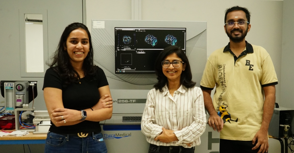
Researchers from the Department of Bioengineering, Indian Institute of Science (IISc), Bengaluru have now developed a biocompatible small molecule that can be used as a minimally invasive contrast agent for Photoacoustic (PA) Tomography. In this method, a near-infrared (NIR) laser beam is shined on light-absorbing molecules (chromophores) sent to the target region, which then expand, creating a pressure change.
The change can be picked up as an auditory signal, and analysing these signals allows scientists to construct 3D images of the target region. The method is particularly useful for pinpointing superficial tumours.
Currently, photoacoustic imaging in a clinical setting only makes use of natural chromophores like haemoglobin, which are already present in the human body. Its oxygenated and deoxygenated states have different optoacoustic signatures that act as useful biomarkers of abnormal metabolic processes.
In the new study, the IISc team designed an external molecule – not typically found in human cells – to provide better sensitivity and contrast, which makes it easier to differentiate the target region from normal tissues. The molecule, referred to as GPc, consists of four glucose units conjugated to a scaffold made of zinc-phthalocyanine. The authors, however, suggest that more studies are needed to fully evaluate GPc’s function once it enters the cells.